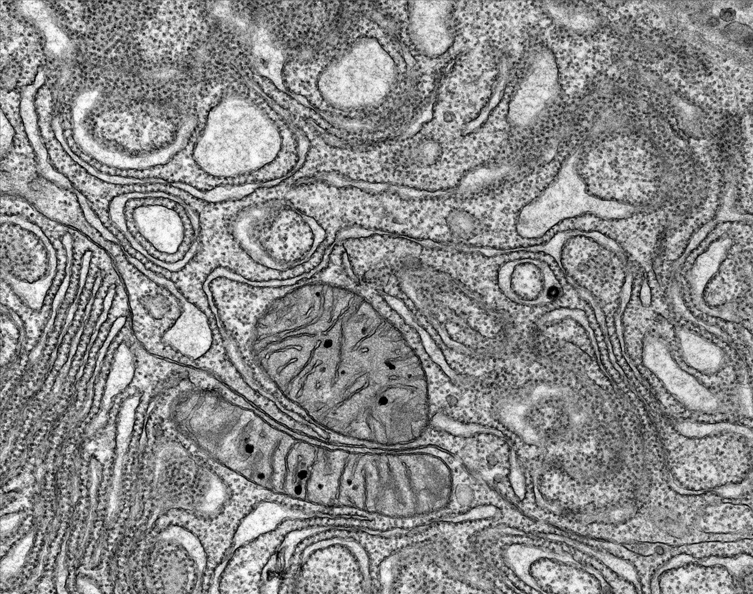
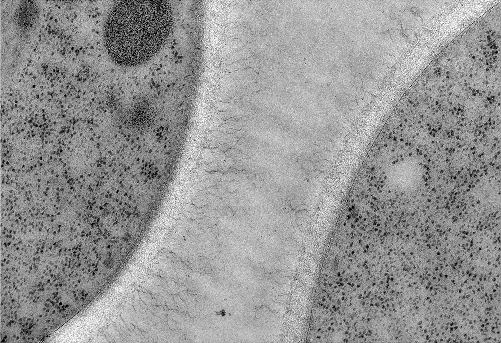
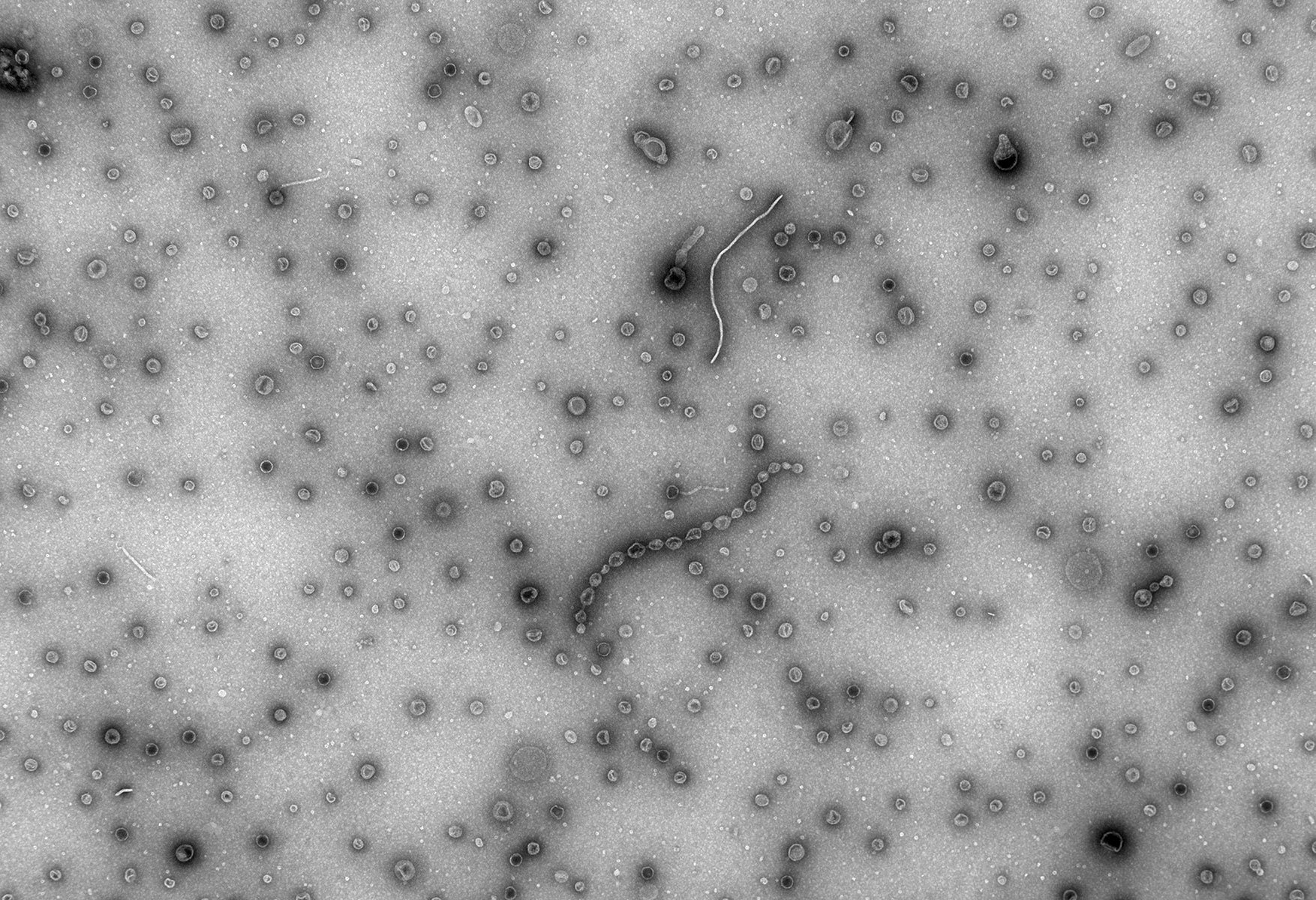
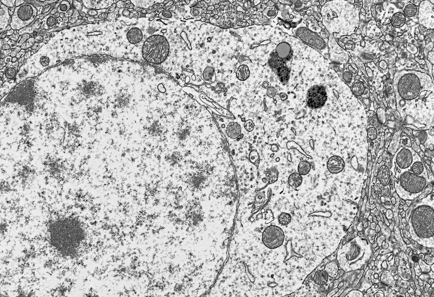
AMT Imaging

TEM Imaging Systems

Adam NS12 Saturday_018
NS43_11
NS43_11
NS43Test_06Feb19_Phage_001
NS43Test_06Feb19_Phage_003
Penn State #1_006
Penn State #1_006
Tom2100F_1500kx
Tom2100F_1500kx
0
Menu
  • External link opens in new tab or window
  • External link opens in new tab or window
  • External link opens in new tab or window
  • External link opens in new tab or window
  • External link opens in new tab or window
  • External link opens in new tab or window

Lens Front

AMT Imaging System Design

Since the invention of the Transmission Electron Microscope (TEM) in the 1930s, this imaging and analytical tool has become the mainstay for examination of structures, beyond the resolving power of light-based microscopes. Found in laboratories of virtually all disciplines, the recording of images has evolved from film to digital formats.  With the full spectrum of applications and available TEM manufacturers and models, there must be a complete range of camera designs and mounting locations, in order to satisfy all requirements and budgets. AMT provides the highest performance optics, by specially designing finite-conjugate lenses for all our products. This feature has allowed AMT to continually take advantage of improvements in sensor technology, geometries, and electronics. We are proud to have pioneered the use of high-resolution RES™ phosphors, which minimize electron scattering effects and provide superior sensitivity.


AMT offers a complete line of digital imaging capabilities from 4-megapixel to 43-megapixel formats. All systems provide sharp images, with high dynamic range and low noise, and AMT provides choices of side-, mid- or low-mount locations depending on customer requirements.

Unique Technology

(Click the active circles to read more)

The scientific grade sensors used in AMT's cameras are customized specifically for TEM applications. Cameras either have thermo-electric cooling or run at ambient temperature. All have negligible thermal noise levels at TEM exposure intervals. These cameras have distinctive advantages in readout speed and image quality compared to competing systems.

X

AMT has enhanced laboratory efficiency and productivity by using the latest innovations in fast readout, low noise imaging sensors, and designing an optical, mechanical, and software interface exclusively for TEM users. Custom lenses, specifically designed for this application, offer outstanding resolution and sensitivity compared to alternate optical designs. The exceptionally high MTF and high sensitivity of these lenses are key reasons for AMT's unmatched performance and, unlike competitive fiber optic-based systems, allow the optimized and efficient sampling of today’s smaller sensor geometries.

X

Performance is enhanced further by AMT's unique and proprietary phosphor designs. Balancing phosphor size, thickness, and grain size for specific sensors and not limited by fiber optic dimensions, allows AMT to optimize signal and resolution for each camera and TEM mounting geometry.

X


Camera Locations


Camera location on the microscope column physically determines the field-of-view (FOV) and “magnification factor” with respect to the microscope’s read-out magnification. The wide-angle side-mount option is popular for the large viewing area and ease of surveying samples. Current 5- and 12-Megapixel cameras allow for digital magnification post acquisition, and these are the most cost-effective solutions for general purpose use. AMT’s pioneering of the mid-mount design locates the camera at the optimum position to minimize any TEM image distortion and takes advantage of the larger format sensors that are now available. Finally, for the highest magnification and resolution requirements, the low-mount position enables microscopists to capture the ultimate in fine structure images.
 
Optimum microscope/camera configurations are defined not only by application, but by the best suited position on the microscope for a specific application.

Camera Locations


Side-Mount Cameras

  • Mounting: Within the 35mm film camera "side" position
  • Mounting Attribute: Widest viewing area and highest apparent sensitivity
  • Applications: Biology, Pathology
  • Camera Format: 4Mpix --> 12Mpix
 

Side Mount Description

The Side-Mount systems are positioned perpendicular to the optical axis of the microscope, in the area typically reserved for the 35mm film camera, and well above the fluorescent viewing screen. This configuration provides the widest viewing area and highest apparent sensitivity. The field-of-view (FOV) matches or exceeds plate film. In operation, the electron beam interacts with the phosphor-coated scintillator, projecting an image to a mirror, which in turn projects the image to the optical lens/sensor assembly located outside the vacuum. The scintillator mechanics retract to allow full use of the normal viewing screen. These systems are ideal for typical work in biology and clinical pathology, where context is extremely critical at lower magnifications, coupled with middle to high magnifications used for applications, such as virology. The side-mount systems are also useful for diffraction work where a large field-of-view is advantageous.

Mid-Mount Cameras

  • Mounting: Directly beneath TEM fluorescent viewing screen
  • Mounting Attribute: Extremely sharp image with no TEM-related image distortion
  • Applications: Biology, Pathology, Materials, Tomography
  • Camera Format: 5Mpix --> 43Mpix
 

Mid Mount Description

Pioneered by AMT, the Mid-Mount configurations are positioned on-axis in direct line with the electron beam. Cameras mounted in this position are at or close to the level of the traditional film/plate camera and can be mounted directly beneath the fluorescent viewing screen. Taking advantage of this optimized film position, the Mid-Mount has virtually no TEM-related image distortion. This location supports a large field-of-view (FOV) while still allowing the TEM to reach lattice resolution. The Mid-Mount phosphors can be physically larger than the Side-Mounted phosphor, which in turn results in larger phosphor "pixels" and better optical quality. The greatest benefit of the Mid-Mount camera position is that it optimizes the microscope manufacturer's design for distortion-free documentation. In addition, the stated microscope magnification is similar to the image at the sensor.

Low-Mount Cameras

  • Mounting: Below the TEM film/plate
  • Mounting Attribute: Highest magnification factors at maximum resolution with 10 times or more magnification than the TEM states
  • Applications: Materials Science, In-Situ Studies
  • Camera Format: 5Mpix --> 43Mpix


Low Mount Description

Low-Mount cameras are mounted on-axis in direct line with the electron beam, are mounted below the traditional film/plate camera, and have the highest magnification factors at maximum resolution. With magnifications of 10 times or more beyond the stated microscope magnification, this position is favorable for work typical of materials research.

AMT Imaging Software

Since inception, AMT has dedicated its software efforts toward innovating based on direct input from the more than 2000 systems it has deployed worldwide. In doing so, AMT has developed a user interface that provides sophisticated capability while still maintaining operational simplicity. The modular architecture segments image acquisition from analysis and provides seamless acquisition, image processing, and image storage.


Whether one is using live image survey or viewing the image post-acquisition, the desktop environment features the prominent display of the user's specimen image. Segmenting the screen to display camera controls to the right side of the image, pull-down menu tabs for infrequently used commands and user-specific imaging tools on the left side, keeps workflow simple. For more detailed explanations of software and operation, please see the AMT Camera Manual link below.
External link opens in new tab or windowAMT Camera ManualAMT Camera Manual

AMT Image Capture Engine Screenshot

Column

UPCOMING EVENTS

M&M2025
July 27th - 31st
M&M July 28th - August 1st 2024


Visit Us This Year in Booth 1417


Column

CONTACT INFORMATION


Mailing Address: 242 W Cummings Park, Woburn, MA 01801
Office and Sale Phone: External link opens in new tab or window(978) 774-5550
Fax: (978) 739-4313
Office and Sales Email: inquiry@amtimaging.com
Service Phone: External link opens in new tab or window(781) 226-8382
Service Email: support@amtimaging.com

Home | Key Technology | General Imaging | Cryo Imaging | About | News | Careers | Contact

close lightbox